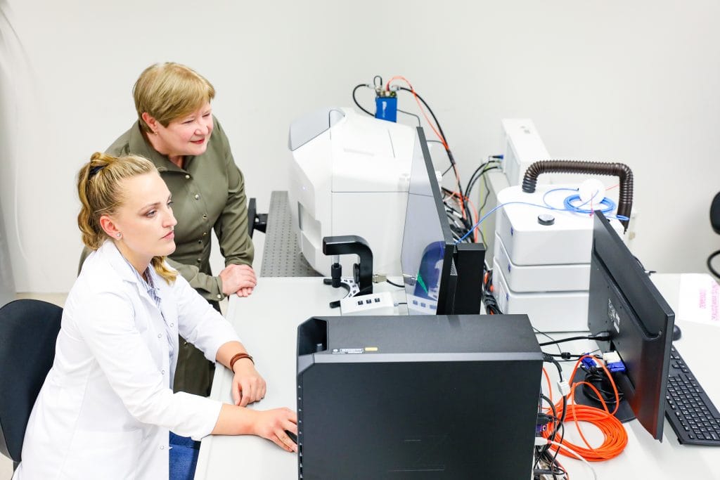
Advanced imaging technology is a novelty in the Baltic region, and such a microscope is the only one. Research, which will be carried out using advanced laboratory equipment, will contribute to new scientific discoveries that will help to improve the level of biomedical sciences in Lithuania and bring it closer to European standards.
“Lithuania is taking the lead in the field of biomolecular sciences, and the technological equipment in Kaunas is expected to become a pivot not only for research but also for further technological development,” says Prof. Dr Vilmantė Borutaitė, chief researcher and the head of the Biochemistry Laboratory of the LSMU Institute of Neuroscience.
Revolutionary speed and detail
The light sheet fluorescence microscope of Internationally recognised German manufacturer ZEISS Microscopy allows life science and biomedical professionals to explore live samples.

“The advantage of this microscope is that it combines speed and high resolution simultaneously, which is why it is successfully used and will be widely applied in monitoring and evaluating fast processes for which the speed of other microscopy techniques was insufficient. On the other hand, it provides an opportunity to scan a large volume very quickly, which allows us to analyse the structure of the entire tissue,” says Urtė Neniškytė, a neuroscientist at the VU Life Sciences Center.
A high-speed digital video camera captures a sample stretch in milliseconds. It holds millions of pixels, and such stretches are recorded in seconds. “By using conventional confocal microscopy, we can photograph one cell only, whereas this microscope allows us to photograph all the cells in that tissue sample in just a few seconds and gather significantly more information,” the neuroscientist continues.
According to Urtė Neniškytė, faster visualisation helps to prevent phototoxicity. Due to quicker imaging, the sample is shorter exposed to laser light, which is intense and toxic to living tissues.
Will help find more effective treatments for cancer

According to V. Borutaitė, it is planned that the new equipment will be used to study spatial changes in nervous system cells in models of neurodegenerative diseases. New drugs will be sought, and diagnostic and treatment methods will be developed based on the knowledge gained from three-dimensional model systems of brain cells. “Another area of research is neurooncology, where the application of the new research methodology will make it possible to understand the patterns of malignant tumours better and, once understood, to search for more effective treatment methods,” says the professor.
The new microscope will also help researchers of LUHS and other institutions working in regenerative medicine, who are developing methods for the biological reconstruction of the skin, joints, other organs, and tissues using adult human tissue stem cells and other methods. Advanced technology will allow a more comprehensive application of imaging and research methodologies of spatial cellular systems, adapting them to research into the pathogenesis of various diseases. This means opening up new and unique applications for advancing biomedical research.
“It is the implementation of innovations in the field of biomedicine that opens new opportunities for Lithuania and creates added value for the entire biomedical research system. We are glad that we were able to offer an innovative solution to Lithuanian researchers that meets world-class standards.” Says Dainius Vasiliauskas, director of “Inospectra”, a company that provides laboratory and analytical equipment solutions.
Ongoing staff training
The specialists of the company Inospectra took care of the logistics of the new equipment, prepared it for work, and continued to work closely with the scientists, providing operational service. Most importantly, they ensure proper training for workers in laboratory use of advanced technology.

“The first training left a good impression and contributed to the generation of new thoughts and ideas, and at the same time raised new tasks: take over the advantages of this technology at best and use it to the maximum for future research and work,” says Silvija Jankevičiūtė, a researcher at the LSMU Institute of Neuroscience, who, along with other scientists, was the first to test the new light sheet microscope.
Lithuanian University of Health Sciences (LUHS), together with Vilnius University Life Sciences Centre (VU GMC), is implementing the infrastructure improvement project “Centre for Computational, Structural, and Systems Biology – CossyBio”, the total value of which is 6.18 million euros. This project combines and develops state-of-the-art laboratory equipment for cellular imaging systems and molecular structures, monitoring biomolecular processes, and research in living organisms.

